Digestion is a round-the-clock control problem. Food has to be mixed, moved along at the right speed, met with the right secretions at the right moment, and monitored continuously across several metres of tube — all of it without a single conscious instruction, whether you are awake, asleep, or thinking about something else entirely.
The obvious candidate for that job is the brain. But the brain is a long way from the intestine, and it has other things to do. The body's solution is more radical: a second nervous system, built directly into the wall of the gut, that lets the organ largely run itself.
That system is called the enteric nervous system, and it is not a figure of speech. It is made of real neurons — hundreds of millions of them — wired into complete working circuits. This article is about where it sits, the elegant experiment from 1899 that proved it works on its own, and why it has changed the way medicine talks about digestive symptoms.
A nervous system hidden in a tube
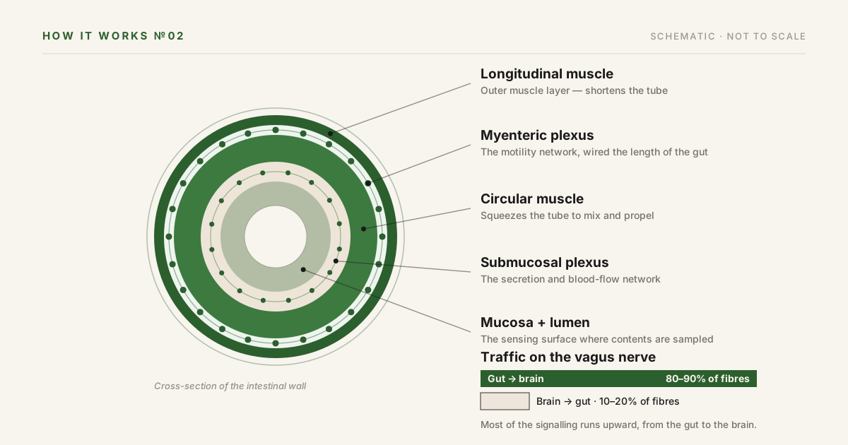
The wall of the digestive tract is built in layers: an outer sheet of muscle running lengthwise, an inner sheet wrapping around the tube, then a connective-tissue layer, and finally the mucosa — the lining that faces the contents. The enteric nervous system lives in two thin sheets tucked between those layers, shown as the dotted rings in the diagram above.
The human version contains somewhere between 200 and 600 million neurons, gathered into many thousands of small clusters called ganglia.1 Most of them belong to two networks: the myenteric plexus, sandwiched between the two muscle layers, and the submucosal plexus, just beneath the lining. The myenteric plexus is not a local feature — it forms one continuous net from the upper esophagus all the way to the internal anal sphincter.1
What makes this a nervous system rather than mere wiring is that the circuits are complete. The gut wall contains its own sensory neurons, its own interneurons to process the signal, and its own motor neurons to act — controlling the muscle, the movement of fluid across the lining, and local blood flow, without waiting for the brain's permission.1
The 1899 experiment that proved it
In 1899, two London physiologists, William Bayliss and Ernest Starling — the pair who would go on to discover the hormone secretin — asked a simple question: what does the intestine do when something presses on it from the inside?
Working with anaesthetized animals, they found that the answer has a beautiful shape. Pressure at any point makes the muscle just behind that point contract, and the muscle just ahead of it relax. The result is a travelling wave that pushes the contents onward — always onward — no matter where the stimulus lands. They called it “the law of the intestine,” and they showed that it is organised by what they carefully named the local nervous mechanism: the nerves in the wall itself.2
The modern verdict goes further. Cutting the nerve connections between the gut and the central nervous system has surprisingly minor effects on digestion; the small intestine and colon carry on, run by their onboard circuits.1 The statement has limits — the esophagus and stomach lean much more heavily on the brain, and defecation is coordinated from centres in the spinal cord1 — but on its home territory, the gut is strikingly self-governing.
The line to the brain runs mostly upward
None of this means the gut is cut off. It is connected — mainly through the vagus nerve, the long wandering nerve that links the brainstem to the organs of the chest and abdomen. What is surprising is the direction of the traffic.
Of the fibres in the vagus, roughly 80 to 90 percent are afferent: they carry information up, from the gut to the brain. Only 10 to 20 percent carry commands down.3 On this connection, in other words, the brain spends far more of its time listening than instructing.
And there is plenty to listen to. The vagal endings in the digestive tract include stretch receptors, tension receptors and chemical sensors, reporting on what has arrived and how the wall is responding.3 The everyday phrase “gut feeling” is not anatomy, but it points at something real: a dense, constant stream of reports flowing from the digestive tract to the brain, most of which never reaches conscious attention.
Most of your serotonin is not in your head
Serotonin is best known as a brain chemical. Yet roughly 95 percent of the body's serotonin is found in the gut, released by specialised cells in the lining called enterochromaffin cells.4 There it works as a local signalling molecule — among other things, helping to regulate the motility of the bowel.4
That single number does a lot of quiet work. It is a reminder that molecules we file under “mood” are also — and in sheer quantity, mainly — digestive molecules, and that the chemistry of the gut and the chemistry of the nervous system were never separate subjects to begin with.
What this means if you are the patient
For a long time, digestive symptoms without visible damage — no ulcer, no inflammation, nothing abnormal to see — sat in an awkward category often labelled “functional.” The field has moved. The current international classification, Rome IV, formally describes these conditions as disorders of gut–brain interaction: problems that can arise in the signalling between the two nervous systems — in motility, in the sensitivity of the gut's own sensors, in how the brain processes what the gut reports — rather than in the visible structure of the organ.5 They are among the most common diagnoses in gastroenterology.5
Knowing this machinery exists explains a few things people notice:
- Why digestion carries on, day and night, with no conscious effort at all
- Why emotions and stress can register so distinctly in the digestive tract
- Why a symptom can be entirely real even when the organ looks structurally normal
What any of this means for one person's symptoms is a clinical question, and it belongs to the clinician who knows the case. If this article leaves you with a question about your own digestion, that is exactly the question to bring to your family physician, nurse practitioner or specialist.
Dr. Stephane Gauthier provides gastroenterology care in North Bay and serves patients across Northern Ontario. His practice includes inflammatory bowel disease, colonoscopy and advanced endoscopy, liver disease, GERD and esophageal conditions, and internal medicine. He accepts patients by physician referral.
General information only. This article describes an area of human physiology. It is not medical advice, it is not a diagnosis, and it does not recommend any test or treatment. It cannot account for your individual circumstances. For guidance about your own health, speak with your family physician, nurse practitioner or specialist.
La digestion est un problème de contrôle qui ne s'arrête jamais. Il faut mélanger les aliments, les faire avancer à la bonne vitesse, y ajouter les bonnes sécrétions au bon moment et surveiller en continu plusieurs mètres de tube — le tout sans la moindre instruction consciente, que vous soyez éveillé, endormi ou occupé à penser à tout autre chose.
Le candidat évident pour cette tâche est le cerveau. Mais le cerveau est loin de l'intestin, et il a d'autres occupations. La solution du corps est plus radicale : un deuxième système nerveux, construit directement dans la paroi du tube digestif, qui permet à l'organe de se gouverner en grande partie lui-même.
Ce système s'appelle le système nerveux entérique, et ce n'est pas une figure de style. Il est fait de vrais neurones — des centaines de millions — reliés en circuits complets et fonctionnels. Cet article porte sur son emplacement, sur l'élégante expérience de 1899 qui a prouvé qu'il fonctionne par lui-même, et sur la façon dont il a changé la manière dont la médecine parle des symptômes digestifs.
Un système nerveux caché dans un tube
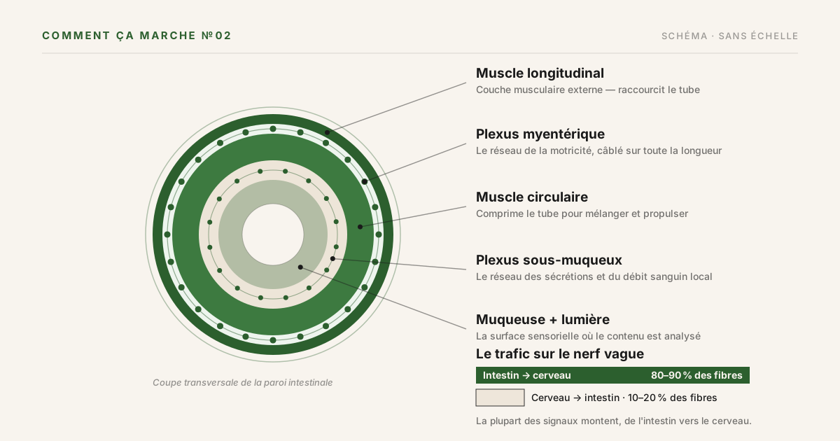
La paroi du tube digestif est construite en couches : une nappe musculaire externe orientée dans le sens de la longueur, une nappe interne qui enserre le tube, puis une couche de tissu conjonctif et enfin la muqueuse — le revêtement qui fait face au contenu. Le système nerveux entérique loge dans deux minces feuillets glissés entre ces couches, représentés par les anneaux pointillés du schéma ci-dessus.
Chez l'humain, il contient entre 200 et 600 millions de neurones, regroupés en plusieurs milliers de petits amas appelés ganglions.1 La plupart appartiennent à deux réseaux : le plexus myentérique, pris en sandwich entre les deux couches musculaires, et le plexus sous-muqueux, juste sous le revêtement. Le plexus myentérique n'est pas une structure locale : il forme un filet continu depuis le haut de l'œsophage jusqu'au sphincter anal interne.1
Ce qui en fait un système nerveux plutôt qu'un simple câblage, c'est que les circuits sont complets. La paroi intestinale contient ses propres neurones sensoriels, ses propres interneurones pour traiter le signal et ses propres neurones moteurs pour agir — contrôlant le muscle, les mouvements de liquide à travers le revêtement et le débit sanguin local, sans attendre la permission du cerveau.1
L'expérience de 1899 qui l'a prouvé
En 1899, deux physiologistes londoniens, William Bayliss et Ernest Starling — ceux-là mêmes qui découvriraient ensuite l'hormone sécrétine — se sont posé une question simple : que fait l'intestin quand quelque chose appuie sur lui de l'intérieur ?
En travaillant sur des animaux anesthésiés, ils ont découvert que la réponse a une forme magnifique. Une pression en un point quelconque fait se contracter le muscle situé juste en amont et se relâcher celui situé juste en aval. Le résultat est une onde qui pousse le contenu vers l'avant — toujours vers l'avant — peu importe où tombe le stimulus. Ils ont appelé cela « la loi de l'intestin » et ont montré qu'elle est organisée par ce qu'ils ont soigneusement nommé le mécanisme nerveux local : les nerfs de la paroi elle-même.2
Le verdict moderne va plus loin. Couper les connexions nerveuses entre l'intestin et le système nerveux central n'a que des effets étonnamment mineurs sur la digestion ; l'intestin grêle et le côlon continuent, pilotés par leurs circuits embarqués.1 L'affirmation a ses limites — l'œsophage et l'estomac dépendent beaucoup plus du cerveau, et la défécation est coordonnée depuis des centres de la moelle épinière1 — mais sur son propre territoire, l'intestin est remarquablement autonome.
La ligne vers le cerveau monte, surtout
Rien de tout cela ne signifie que l'intestin est isolé. Il est connecté — principalement par le nerf vague, ce long nerf « vagabond » qui relie le tronc cérébral aux organes du thorax et de l'abdomen. Ce qui surprend, c'est le sens de la circulation.
Parmi les fibres du nerf vague, environ 80 à 90 % sont afférentes : elles transportent l'information vers le haut, de l'intestin vers le cerveau. Seulement 10 à 20 % transmettent des commandes vers le bas.3 Sur cette liaison, autrement dit, le cerveau passe beaucoup plus de temps à écouter qu'à donner des ordres.
Et il y a de quoi écouter. Les terminaisons vagales du tube digestif comprennent des récepteurs d'étirement, des récepteurs de tension et des capteurs chimiques, qui rendent compte de ce qui est arrivé et de la réaction de la paroi.3 L'expression courante « se fier à ses tripes » n'est pas de l'anatomie, mais elle pointe vers quelque chose de réel : un flux dense et constant de rapports circulant du tube digestif vers le cerveau, dont la majeure partie n'atteint jamais l'attention consciente.
La majorité de votre sérotonine n'est pas dans votre tête
La sérotonine est surtout connue comme une molécule du cerveau. Pourtant, environ 95 % de la sérotonine du corps se trouve dans l'intestin, libérée par des cellules spécialisées du revêtement appelées cellules entérochromaffines.4 Elle y agit comme molécule de signalisation locale — contribuant, entre autres, à réguler la motricité de l'intestin.4
Ce simple chiffre accomplit beaucoup de travail discret. Il rappelle que des molécules que nous classons sous « humeur » sont aussi — et, en quantité pure, surtout — des molécules digestives, et que la chimie de l'intestin et celle du système nerveux n'ont jamais été des sujets séparés.
Ce que cela signifie si vous êtes le patient
Pendant longtemps, les symptômes digestifs sans lésion visible — pas d'ulcère, pas d'inflammation, rien d'anormal à voir — sont restés dans une catégorie inconfortable, souvent étiquetée « fonctionnelle ». Le domaine a évolué. La classification internationale actuelle, Rome IV, décrit désormais formellement ces affections comme des troubles de l'interaction intestin-cerveau : des problèmes qui peuvent naître dans la signalisation entre les deux systèmes nerveux — dans la motricité, dans la sensibilité des capteurs propres à l'intestin, dans la façon dont le cerveau traite ce que l'intestin rapporte — plutôt que dans la structure visible de l'organe.5 Ces diagnostics comptent parmi les plus fréquents en gastroentérologie.5
Savoir que cette machinerie existe explique certaines choses que l'on remarque :
- Pourquoi la digestion se poursuit, jour et nuit, sans le moindre effort conscient
- Pourquoi les émotions et le stress peuvent se faire sentir si distinctement dans le tube digestif
- Pourquoi un symptôme peut être parfaitement réel même quand l'organe paraît structurellement normal
Ce que tout cela signifie pour les symptômes d'une personne en particulier est une question clinique, qui appartient au clinicien qui connaît le dossier. Si cet article vous laisse avec une question sur votre propre digestion, c'est précisément la question à poser à votre médecin de famille, à votre infirmière praticienne ou à votre spécialiste.
Le Dr Stephane Gauthier offre des soins en gastroentérologie à North Bay et dessert des patients partout dans le Nord de l'Ontario. Sa pratique comprend les maladies inflammatoires de l'intestin, la coloscopie et l'endoscopie avancée, les maladies du foie, le RGO et les affections de l'œsophage, ainsi que la médecine interne. Il accepte les patients sur référence médicale.
Information générale seulement. Cet article décrit un domaine de la physiologie humaine. Il ne constitue pas un avis médical, il ne pose aucun diagnostic et ne recommande aucun examen ni traitement. Il ne peut pas tenir compte de votre situation personnelle. Pour des conseils sur votre propre santé, consultez votre médecin de famille, votre infirmière praticienne ou votre spécialiste.
Sources Sources
- Furness JB, Callaghan BP, Rivera LR, Cho HJ. “The enteric nervous system and gastrointestinal innervation: integrated local and central control.” Advances in Experimental Medicine and Biology, 2014;817:39–71. PMID 24997029 · doi:10.1007/978-1-4939-0897-4_3
- Bayliss WM, Starling EH. “The movements and innervation of the small intestine.” The Journal of Physiology, 1899;24(2):99–143. PMC1516636 · doi:10.1113/jphysiol.1899.sp000752
- Breit S, Kupferberg A, Rogler G, Hasler G. “Vagus Nerve as Modulator of the Brain–Gut Axis in Psychiatric and Inflammatory Disorders.” Frontiers in Psychiatry, 2018;9:44. PMC5859128 · doi:10.3389/fpsyt.2018.00044
- Berger M, Gray JA, Roth BL. “The expanded biology of serotonin.” Annual Review of Medicine, 2009;60:355–366. PMC5864293 · doi:10.1146/annurev.med.60.042307.110802
- Drossman DA. “Functional Gastrointestinal Disorders: History, Pathophysiology, Clinical Features and Rome IV.” Gastroenterology, 2016. PMID 27144617 · doi:10.1053/j.gastro.2016.02.032